
(Mentors: Laura Anne Lowery, Assistant Prof. of Biology; Ken Burch, Associate Prof. of Physics)
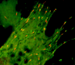
Description: This project is focused on the fundamental question of how neuronal growth cones are accurately and precisely guided to their targets during development. Understanding these growth dynamics is ultimately crucial for brain development. The mechanisms by which cytoskeletal dynamics bring about specific changes in growth cone microtubule dynamics are unresolved. We hypothesize that spatial changes in microtubule regulators occur during growth cone steering. Testing such a hypothesis requires simultaneous real-time, in-situ imaging of the numerous possible regulators, which is not possible with typical fluorescence microscopy, where only two to four regulators can be imaged (see Figure 1). To overcome this difficulty, we will focus on the feasibility of Raman microscopy to simultaneously image the levels of numerous microtubule regulators (such as tubulin and TACC3). Specifically, the student in the Lowery lab will prepare neuronal growth cones and perform initial optical/fluorescence measurements, while the student in the Burch group will optimize the existing Raman microscope to image live cells by measuring the resulting vibrational spectra. The two students will collaborate on developing sample holders compatible with neuronal growth cones that simultaneously improve the Raman signal levels (via Fabry-Perot Interference). The students will also collaborate on analyzing the Raman spectra and comparing images obtained by the two different methods (Raman versus fluorescence)